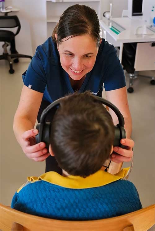

A tympanometry test measures how well your eardrum moves in response to changes in air pressure, giving us a snapshot of what’s happening in your middle ear space: the air-filled cavity behind the drum that houses the tiny bones responsible for conducting sound. It takes about a minute per ear, requires no response from you, and often reveals problems that don’t show up on a standard hearing test. It’s common for someone to assume their hearing loss must be nerve damage or age-related, only for a tympanogram to reveal fluid sitting quietly behind an intact eardrum, sometimes for months.
This isn’t a hearing test in the traditional sense. You won’t be pressing buttons or repeating words. Tympanometry measures mechanics: whether the structures designed to transmit sound into your inner ear are moving the way they should, or whether something (fluid, negative pressure, a hole, scarring) is getting in the way.
What Tympanometry Actually Measures
Your eardrum should move. That sounds obvious, but the amount it moves, and when it moves best, tells us an enormous amount about middle ear health.
During the test, a soft probe tip seals your ear canal and generates three things simultaneously: a constant low-pitched tone (usually 226 Hz in adults; a higher 1000 Hz tone is used for young babies), varying air pressure (from positive to negative), and a microphone measuring how much sound bounces back. When your eardrum is stiff, because there’s pressure behind it, or fluid, or a perforation, it doesn’t absorb sound efficiently, so more gets reflected. When it moves freely, less sound bounces back. The tympanogram plots eardrum mobility (technically called admittance or compliance) against pressure.
The pressure component is crucial. In a healthy middle ear, the air pressure on both sides of the eardrum is equal: atmospheric pressure in the ear canal, the same pressure in the middle ear space (maintained via the Eustachian tube connecting to the back of your nose). The eardrum moves best when those pressures match. If your middle ear pressure has dropped, common with a blocked Eustachian tube, the eardrum moves best at negative pressure. If there’s fluid filling the space, the drum barely moves at all, regardless of pressure.
This is graphed in real time. The peak of the curve shows us where your eardrum is most compliant, and the height and shape of that peak tells us whether the middle ear system is functioning normally.
The Three Tympanogram Types You’ll Actually Encounter
Tympanometry classification uses a lettering system (Type A, B, C and a few sub-types) that has been standard since James Jerger described it in 1970. It’s held up because it’s clinically useful, not because audiologists love arbitrary categories.
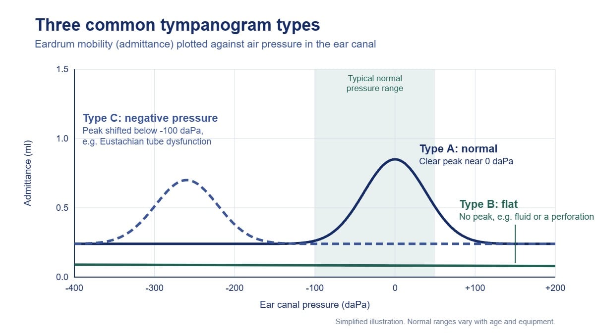
Type A is normal. The peak sits between +50 and -100 daPa (decaPascals, the pressure unit), meaning your middle ear pressure is close to atmospheric. The peak is sharp and well-defined, indicating the eardrum is moving freely. Compliance falls within normal range, typically 0.3 to 1.5 ml (or cm³) for adults. You’ll sometimes see sub-types: Type As indicates reduced compliance: a stiff system, seen with otosclerosis or tympanosclerosis. Type Ad shows excessive compliance, sometimes present with ossicular discontinuity or a very flaccid eardrum. Most of the time, a Type A trace means middle ear function isn’t the cause of your hearing problem, if you have one.
Type B is flat. No discernible peak, minimal change in admittance across the entire pressure sweep. This is the tympanogram I see most often when something’s genuinely wrong. The classic cause is middle ear effusion: fluid (serous or mucoid) filling the space behind the eardrum, preventing it from moving. It’s incredibly common in children post-upper respiratory infection, but adults get it too, especially after flying with a head cold or in the setting of chronic Eustachian tube dysfunction. The other major cause of a Type B trace is a tympanic membrane perforation, which usually shows up with a larger than expected ear canal volume reading. If there’s a hole in the drum, pressurising the ear canal doesn’t create a pressure differential across the membrane, air just moves through the perforation, so you get a flat line. Clinically, I can usually differentiate these with otoscopy (looking in the ear), but the tympanogram itself won’t tell you which it is. Both result in conductive hearing loss, but the management is completely different. A Type B result can also occur when wax completely blocks the ear canal, in which case the volume reading is unusually small.
Type C shows a distinct peak, but it’s shifted into negative pressure territory, typically beyond -100 daPa, sometimes as far as -300 or -400 daPa. This indicates Eustachian tube dysfunction: the middle ear has become a sealed chamber with reduced pressure, often because the Eustachian tube isn’t opening properly to equalise with atmospheric pressure. The eardrum gets sucked inward (you can often see this retraction on otoscopy), and it moves best when we recreate that negative environment in the ear canal. Type C tympanograms are common with nasal congestion, allergies or Eustachian tube dysfunction. Sometimes they resolve spontaneously once the cold clears. Sometimes they progress to Type B if fluid gets pulled into the low-pressure space. A Type C trace usually correlates with mild conductive hearing loss and a sensation of fullness or muffled hearing, though not always.
How Tympanometry Fits Into Diagnostic Audiometry
I run tympanometry on nearly every patient, regardless of their presenting complaint, because it takes 90 seconds and rules out or confirms an entire category of pathology. It’s part of the immittance test battery, which also includes acoustic reflex testing (measuring the stapedius muscle contraction in response to loud sound: useful for differential diagnosis, but that’s another article).
Tympanometry complements pure tone audiometry. If your hearing test shows a conductive loss (an air-bone gap, meaning sound delivered through headphones is heard less well than sound delivered by bone conduction, which bypasses the middle ear), I expect an abnormal tympanogram. If I see a 30 dB air-bone gap and a perfect Type A tympanogram, I’m rechecking everything, because those findings don’t fit together. Conversely, you can have a completely normal audiogram and still have tympanometric abnormalities, especially early or resolving middle ear issues that haven’t yet shifted hearing thresholds enough to appear on the chart.
It’s also invaluable before hearing aid fitting. If someone presents wanting hearing aids and I find a Type B tympanogram with conductive loss, they’re going to their GP or an ENT specialist first, not walking out with devices. You don’t amplify a medical problem; you treat it.
What to Expect During the Test
You’ll sit still while I place a probe tip with a rubber cuff into your ear canal: it needs a seal, so I’ll choose a tip size that fits snugly but comfortably. The machine generates pressure changes; you’ll feel a sensation of fullness, like your ears do on an aeroplane, sometimes accompanied by a crackling or popping. The tone is audible but not loud. The entire procedure is over in one to two minutes per ear.
You don’t need to respond or do anything. Unlike pure tone audiometry, where your accuracy matters, tympanometry is entirely objective. I’m measuring a physical response; you could sleep through it (some kids do) and the results would be identical.
There’s no pain, though if you have an acutely infected or very tender ear, the pressure changes can be briefly uncomfortable. If you have a tympanic membrane perforation, you might feel air moving through the hole: it’s harmless, but let me know if it happens.
When Abnormal Results Matter (and When They Don’t)
Not every Type C tympanogram needs intervention. If you’ve got the tail end of a cold, some negative middle ear pressure is expected and will likely self-resolve. I’ll often retest in two to four weeks if you’re asymptomatic or the hearing loss is minimal.
Type B results, on the other hand, almost always warrant further investigation. Persistent middle ear fluid in an adult, especially unilateral (one-sided), can rarely indicate a nasopharyngeal mass blocking the Eustachian tube opening. It’s uncommon, but it’s the reason we don’t dismiss a flat tympanogram in a 50-year-old as “just a bit of fluid.” I’ll refer for medical assessment.
In children, persistent or recurrent Type B tympanograms, particularly with associated hearing loss, are among the criteria for considering grommets (tympanostomy tubes). That decision involves ENT and often speech pathology if language development is a concern, but the tympanogram provides objective evidence that guides timing and intervention.
The Limits of the Test
Tympanometry won’t diagnose everything. It tells you about the middle ear, not the cochlea, not the auditory nerve, not the brain. A patient with profound sensorineural hearing loss from noise exposure or ageing will likely have a completely normal Type A tympanogram, because their eardrum and ossicles are fine: the problem is deeper.
It also won’t differentiate types of middle ear fluid (serous versus mucoid versus purulent), won’t tell you why the Eustachian tube isn’t working, and won’t identify small perforations that are obscured by debris or wax unless I’m looking with an otoscope as well.
And it’s useless if the ear canal is blocked or the probe seal is poor. A wax-blocked ear can produce an abnormal tympanogram that becomes a normal Type A once the wax is removed. Results should always be read alongside the case history and otoscopy.
If you’ve been told you have an abnormal tympanogram, or if you’re experiencing ear fullness, muffled hearing, or pain, the next step is a full diagnostic assessment, not internet research. At The Audiology Place in Epping and Forestville, tympanometry is included in our comprehensive hearing assessments for adults and children, because understanding where the problem is located is the first step toward actually fixing it.